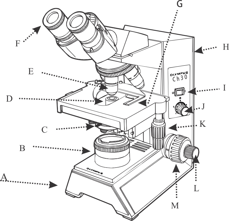
1.Fully label the compound microscope below.

2. What are the ways in which the light can be adjusted on the compound microscope?
3. How is total magnification of the compound microscope determined?
4. Why should you center the image in the visual field before changing power?
5. How is the iris diaphragm opening related to depth of field?
6. What should go over the specimen when you are doing a live mount?
7. Label the drawing below:

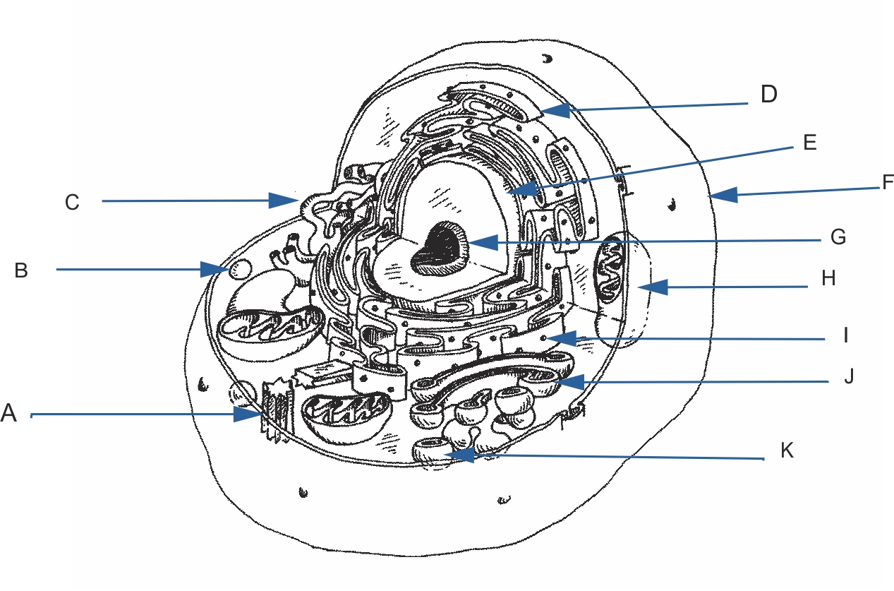
8. Label the eukaryotic cell below:

Give examples of the following:
12. How does a cheek cell different from a plant cell?
13. How does a fungus differ from an algae?
14. How can you make the nucleus visible?
20. Are the cells to the right represent a prokaryotic or eukaryotic organism?
21. Those small round structures within the cells represent what organelles?
|
 |
| 23. The cell on the right represents what group of bacteria? |  |
| 24. This sample stained with iodine shows both chloroplasts and a nucleus, what is its common name? |  |